Keratitis treatment in Alagbole Akute Ajuwon

Overview

Keratitis is an inflammation of the cornea — the clear, dome-shaped tissue on the front of your eye that covers the pupil and iris. Keratitis may or may not be associated with an infection. Noninfectious keratitis can be caused by a relatively minor injury, by wearing your contact lenses too long or by a foreign body in the eye. Infectious keratitis can be caused by bacteria, viruses, fungi and parasites.

If you have eye redness or other symptoms of keratitis, make an appointment to see your doctor. With prompt attention, mild to moderate cases of keratitis can usually be effectively treated without loss of vision. If left untreated, or if an infection is severe, keratitis can lead to serious complications that may permanently damage your vision.

Symptoms

Signs and symptoms of keratitis include:

  • Eye redness
  • Eye pain
  • Excess tears or other discharge from your eye
  • Difficulty opening your eyelid because of pain or irritation
  • Blurred vision
  • Decreased vision
  • Sensitivity to light (photophobia)
  • A feeling that something is in your eye

When to see a doctor

If you notice any of the signs or symptoms of keratitis, make an appointment to see your doctor right away. Delays in diagnosis and treatment of keratitis can lead to serious complications, including blindness.

Causes

Causes of keratitis include:

  • Injury. If any object scratches or injures the surface of your cornea, noninfectious keratitis may result. In addition, an injury may allow microorganisms to gain access to the damaged cornea, causing infectious keratitis.
  • Contaminated contact lenses. Bacteria, fungi or parasites — particularly the microscopic parasite acanthamoeba — may inhabit the surface of a contact lens or contact lens carrying case. The cornea may become contaminated when the lens is in your eye, resulting in infectious keratitis. Over-wearing your contact lenses can cause keratitis, which can become infectious.
  • Viruses. The herpes viruses (herpes simplex and herpes zoster) may cause keratitis.
  • Bacteria. The bacterium that causes gonorrhea can cause keratitis.
  • Contaminated water. Bacteria, fungi and parasites in water — particularly in oceans, rivers, lakes and hot tubs — can enter your eyes when you’re swimming and result in keratitis. However, even if you’re exposed to these bacteria, fungi or parasites, a healthy cornea is unlikely to become infected unless there has been some previous breakdown of the corneal surface — for example, wearing a contact lens too long.

Risk factors

Factors that may increase your risk of keratitis include:

  • Contact lenses. Wearing contact lenses — especially sleeping in the lenses —increases your risk of both infectious and noninfectious keratitis. The risk typically stems from wearing them longer than recommended, improper disinfection or wearing contact lenses while swimming.Keratitis is more common in people who use extended-wear contacts, or wear contacts continuously, than in those who use daily wear contacts and take them out at night.
  • Reduced immunity. If your immune system is compromised due to disease or medications, you’re at higher risk of developing keratitis.
  • Corticosteroids. Use of corticosteroid eyedrops to treat an eye disorder can increase your risk of developing infectious keratitis or worsen existing keratitis.
  • Eye injury. If one of your corneas has been damaged from an injury in the past, you may be more vulnerable to developing keratitis.

Complications

Potential complications of keratitis include:

  • Chronic corneal inflammation and scarring
  • Chronic or recurrent viral infections of your cornea
  • Open sores on your cornea (corneal ulcers)
  • Temporary or permanent reduction in your vision
  • Blindness

Prevention

Caring for your contact lenses

If you wear contact lenses, proper use, cleaning and disinfecting can help prevent keratitis. Follow these tips:

  • Choose daily wear contacts, and take them out before going to sleep.
  • Wash, rinse and dry your hands thoroughly before handling your contacts.
  • Follow your eye care professional’s recommendations for taking care of your lenses.
  • Use only sterile products that are made specifically for contact lens care, and use lens care products made for the type of lenses you wear.
  • Gently rub the lenses during cleaning to enhance the cleaning performance of the contact lens solutions. Avoid rough handling that might cause your lenses to become scratched.
  • Replace your contact lenses as recommended.
  • Replace your contact lens case every three to six months.
  • Discard the solution in the contact lens case each time you disinfect your lenses. Don’t “top off” the old solution that’s already in the case.
  • Don’t wear contact lenses when you go swimming.

Preventing viral outbreaks

Some forms of viral keratitis can’t be completely eliminated. But the following steps may control viral keratitis occurrences:

  • If you have a cold sore or a herpes blister, avoid touching your eyes, your eyelids and the skin around your eyes unless you’ve thoroughly washed your hands.
  • Only use eyedrops that have been prescribed by an eye doctor.
  • Washing your hands frequently prevents viral outbreaks.

SCHEDULE YOUR EYE TEST

Optical eye clinic in alagbole akute ajuwon matogun olambe lambe koye oke iju ishaga fagba
Schedule your eye test with an eye doctor

Astigmatism

According to American Optometric Association (AOA), Astigmatism is a common vision condition that causes blurred vision. It occurs when the cornea (the clear front cover of the eye) is irregularly shaped or sometimes because of the curvature of the lens inside the eye.

An irregularly shaped cornea or lens prevents light from focusing properly on the retina, the light-sensitive surface at the back of the eye. As a result, vision becomes blurred at any distance. This can lead to eye discomfort and headaches.

Astigmatism frequently occurs with other vision conditions like myopia (nearsightedness) and hyperopia (farsightedness). Together these vision conditions are referred to as refractive errors because they affect how the eyes bend or “refract” light.

There are many causes to astigmatism. It can be hereditary and is usually present from birth. It can decrease or increase over time.

A comprehensive optometric examination will include testing for astigmatism. If necessary, your optometrist can provide eyeglasses or contact lenses that correct the astigmatism by altering the way light enters the eyes.

Another option for treating astigmatism is a corneal procedure called orthokeratology (ortho-k). In this painless, noninvasive procedure, the patient wears a series of specially designed rigid contact lenses to gradually reshape the curvature of the cornea.

Laser surgery can also treat some types of astigmatism. The laser changes the shape of the cornea by removing a small amount of eye tissue.

[back to top]

What causes astigmatism?

The curvature of the cornea and lens bends the light entering the eye in order to focus it precisely on the retina at the back of the eye. In astigmatism, the surface of the cornea or lens has a somewhat different curvature.

the surface of the cornea is shaped more like a football instead of round like a basketball, the eye is unable to focus light rays to a single point. Vision becomes out of focus at any distance.

In addition, the curvature of the lens inside the eye can change, resulting in an increase or decrease in astigmatism. This change frequently occurs in adulthood and can precede the development of naturally occurring cataracts.

Sometimes astigmatism may develop following an eye injury or eye surgery.

Astigmatism also occurs due to a relatively rare condition called keratoconus in which the cornea becomes progressively thinner and cone-shaped. This results in a large amount of astigmatism, which causes poor vision that cannot be clearly corrected with eyeglasses. People with keratoconus usually need contact lenses for clear vision and eventually may need a corneal transplant.

An optometrist can diagnose an astigmatism through a comprehensive eye examination. Testing for astigmatism measures how the eyes focus light and determines the power of any optical lenses needed to improve vision. This examination may include:

Visual acuity-When you read letters on a distance chart, you are measuring your visual acuity. Visual acuity is given as a fraction (for example, 20/40). The top number is the standardized testing distance (20 feet) and the bottom number is the smallest letter size read. A person with 20/40 visual acuity would have to get within 20 feet to read a letter that should be seen clearly at 40 feet. Normal distance visual acuity is 20/20.

Keratometry/Topography-A keratometer is the primary instrument used to measure the curvature of the cornea. By focusing a circle of light on the cornea and measuring its reflection, it is possible to determine the exact curvature of that area of the cornea’s surface. This measurement is particularly critical in determining the proper fit for contact lenses. A corneal topographer, which is gaining use, generates a contour map of the cornea and provides even more detail of the cornea’s shape.

Refraction-Using an instrument called a phoropter, your optometrist places a series of lenses in front of your eyes and measures how they focus light. This is performed using a handheld lighted instrument called a retinoscope or an automated instrument that evaluates the approximate focusing power of the eye. Based on your responses, the power is then refined to determine the lenses that allow the clearest vision. Despite improved technology, patient input remains integral in determining vision needs.
With the information from these tests, your optometrist can determine if you have astigmatism. Your optometrist will use these findings, combined with those of other tests performed, to determine the power of any lens correction you need to provide clear, comfortable vision. Once testing is complete, your optometrist can discuss treatment options.

[back to top]

How is astigmatism treated?

People with astigmatism have several options to regain clear vision. They include:

Eyeglasses. People with astigmatism primarily choose eyeglasses to improve their vision. The eyeglasses contain a special cylindrical lens prescription that compensates for the astigmatism. This provides additional power in specific parts of the lens.

Generally, a single-vision lens is prescribed to provide clear vision at all distances. However, patients over age 40 who have presbyopia may need a bifocal or progressive addition lens.

Contact lenses. Some people will have better vision with contact lenses rather than eyeglasses. Contact lenses may provide clearer vision and a wider field of view. However, since contact lenses are worn directly on the eyes, they require regular cleaning and care to safeguard eye health.

Standard soft lenses may not be effective in correcting astigmatism. However, special toric soft contact lenses can correct for many types of astigmatism. Because rigid gas-permeable contact lenses maintain their regular shape while on the cornea, they can compensate for the cornea’s irregular shape and improve vision for people with astigmatism.

Orthokeratology. Orthokeratology (ortho-k) involves the fitting of a series of rigid contact lenses to reshape the cornea. The patient wears contact lenses for limited periods, such as overnight, and then removes them. People with moderate astigmatism may be able to temporarily obtain clear vision without lenses for most of their daily activities. Orthokeratology does not permanently improve vision. If patients stop wearing the retainer lenses, their vision may return to its original condition.

Laser and other refractive surgery procedures. Astigmatism can also be corrected by reshaping the cornea through LASIK (laser in situ keratomileusis) or PRK (photorefractive keratectomy). PRK removes tissue from the superficial and inner layers of the cornea. LASIK removes tissue only from the inner layer of the cornea.
If you have an astigmatism, you have a wide range of options to correct your vision problem. In consultation with your optometrist, you can select the treatment that best meets your visual and lifestyle needs

Hyperopia/hypermetropia (longsightedness or farsightedness): How to correct for it

Hyperopia, or farsightedness, is a common vision problem, affecting about a fourth of the population. People with hyperopia can see distant objects very well, but have difficulty focusing on objects that are up close. The condition is sometimes referred to as “hypermetropia” rather than hyperopia.

Hyperopia symptoms

Farsighted people sometimes have headaches or eye strain and may squint or feel fatigued when performing work at close range. If you get these symptoms while wearing your eyeglasses or contact lenses, you may need an eye exam and a new prescription.

What causes hyperopia/hypermetropia

  1. Signs
  2. Causes
  3. Treatment

hyperopia eyeglasses on top of a book

Schedule an exam

FIND EYE DOCTOR

Hyperopia, or farsightedness, is a common vision problem, affecting about a fourth of the population. People with hyperopia can see distant objects very well, but have difficulty focusing on objects that are up close. The condition is sometimes referred to as “hypermetropia” rather than hyperopia.

Hyperopia symptoms

Farsighted people sometimes have headaches or eye strain and may squint or feel fatigued when performing work at close range. If you get these symptoms while wearing your eyeglasses or contact lenses, you may need an eye exam and a new prescription.

What causes hyperopia/hypermetropia?

Watch this video on what causes blurry vision and how we can correct it.

This vision problem occurs when light rays entering the eye focus behind the retina, rather than directly on it. The eyeball of a farsighted person is shorter than normal.

Many children are born farsighted, and some of them “outgrow” it as the eyeball lengthens with normal growth.

Sometimes people confuse hyperopia with presbyopia, which also causes near vision problems but for different reasons.

Hyperopia treatment

Farsightedness can be corrected with glasses or contact lenses to change the way light rays bend into the eyes.

If your glasses or contact lens prescription begins with plus numbers, like +2.50, you are farsighted.

You may need to wear your glasses or contacts all the time or only when reading, working on a computer or doing other close-up work.

When selecting eyeglasses for the correction of farsightedness, choose aspheric high-index lenses — especially for stronger prescriptions. These lenses are thinner, lighter, and have a slimmer, more attractive profile. Aspheric lenses also reduce the magnified “bug-eye” appearance eyeglasses for hyperopia often cause.

Be aware, though, that high-index aspheric lenses reflect more light than standard plastic lenses. For the best comfort and appearance, make sure the lenses include anti-reflective coating, which eliminates distracting lens reflections.

High-index aspheric lenses for children should be made of lightweight polycarbonate lens material for superior comfort and impact resistance.

Also, photochromic lenses that automatically darken in response to sunlight are highly recommended for kids and anyone who spends a significant amount of time outdoors.

Refractive surgery, such as LASIK or CK, is another option for correcting hyperopia. Surgery may reduce or eliminate your need to wear glasses or contact lenses.

Investigational procedures involving corneal implants may be a future option for correcting hyperopia.

When to see an eye doctor

When to see an Eye-Doctor

1, You always have a headache
2, Your eye is infected
3, There are bright flashes or floaters
4, There is eye pain
5, You have been squinting
6, You have diabetes or it is in the family
7, You have hypertension or it is in the family
8, Excessive light sensitivity
9, Your eyes are always red
10, You spend so much time on computer or phone-screen
11, If you have never had your eyes examined before.

Lagos population distribution

Population distribution of people in Lagos State

Lagos state located in the South West of Nigeria is the smallest state in Nigeria geographically, yet the most populated state in Nigeria. The people who live in Lagos come from different states in Nigeria and are from different ethnic groups in Nigeria.

Most of the people in the below population distribution need eye clinics, eye hospitals, eye care services. They need opticians, optometrists, ophthalmologists for their eye well being.

They also need eye care products/services such as contact lenses, eyeglasses, eye drugs, sports ropes, eye tests, eye treatment, eye surgeries etc.

Eyeupdate eye clinic & optical supplies is committed to providing primary, secondary and tertiary eye care services as well as sales and supplies of optical/ophthalmic equipment and tools.

Eyeupdate clinic & optical supplies is located at 01, Ajuwon junction, Ajuwon bus stop, off Elliot bus stop, Iju-Ishagah, Lagos. You can reach Eyeupdate eye clinic & optical supplies by calling any of these phone numbers: 08107531046, 08034971582

FREE READING GLASSES

GET FREE READING GLASSES ON A SPRING LOADED FRAME

Newly registered patients and residents of Ajuwon, Akute, Alagbole and environs can get free reading glasses every year at Eyeupdate clinic & optical supplies located at 01, Ajuwon junction, Ajuwon bus stop, beside BNPL Filling station.

Information reaching our news room says the free reading glasses initiative is funded as at present solely by Dr. Steven E. N. as part of the “PREVENT BLINDNESS” program. The prevent blindness program is aimed at reducing the preventable causes of blindness in Nigeria.  There is expectation that the gesture will include free eye surgeries in the future. Philanthropists and well meaning individuals and companies are welcomed to become part of this program.

Philanthropists, individuals or companies who may wish to support this initiative can make their donations into the below account :

Bank: Zenith bank
Account number: 1012494861
Account name: Eyeupdate clinic and optical supplies
Contact person : Steven Nwosu
Tel: +2347038125887

OR

Western Union money transfer
Name: Steven Nwosu
Address: 01, Ajuwon junction, Lagos – Nigeria

List of Donors and Amount:

Glaucoma

Glaucoma is a condition that damages your eye‘s optic nerve. It gets worse over time. It’s often linked to a buildup of pressure inside your eyeGlaucoma tends to run in families. You usually don’t get it until later in life.

The increased pressure in your eye, called intraocular pressure, can damage your optic nerve, which sends images to your brain. If the damage worsens, glaucoma can cause permanent vision loss or even total blindness within a few years.

Most people with glaucoma have no early symptoms or pain. Visit your eye doctor regularly so they can diagnose and treat glaucoma before you have long-term vision loss.

If you lose vision, it can’t be brought back. But lowering eye pressure can help you keep the sight you have. Most people with glaucoma who follow their treatment plan and have regular eye exams are able to keep their vision.

Glaucoma Causes

The fluid inside your eye, called aqueous humor, usually flows out of your eye through a mesh-like channel. If this channel gets blocked, the liquid builds up. Sometimes, experts don’t know what causes this blockage. But it can be inherited, meaning it’s passed from parents to children.

Less-common causes of glaucoma include a blunt or chemical injury to your eye, severe eye infection, blocked blood vessels inside your eye, and inflammatory conditions. It’s rare, but eye surgery to correct another condition can sometimes bring it on. It usually affects both eyes, but it may be worse in one than the other.

CONTINUE READING BELOW

Glaucoma Risk Factors

It mostly affects adults over 40, but young adults, children, and even infants can have it. African Americans tend to get it more often, when they’re younger, and with more vision loss.

You’re more likely to get it if you:

  • Are of African American, Irish, Russian, Japanese, Hispanic, Inuit, or Scandinavian descent
  • Are over 40
  • Have a family history of glaucoma
  • Have poor vision
  • Have diabetes
  • Take certain steroid medications such as prednisone
  • Have had an injury to your eye or eyes
  • Have corneas that are thinner than usual
  • Have high blood pressure, heart disease, diabetes, or sickle cell anemia
  • Have high eye pressure
  • Are nearsighted or farsighted

Types of Glaucoma

There are two main kinds:

Open-angle glaucoma. This is the most common type. Your doctor may also call it wide-angle glaucoma. The drain structure in your eye (called the trabecular meshwork) looks fine, but fluid doesn’t flow out like it should.

Angle-closure glaucoma. This is more common in Asia. You may also hear it called acute or chronic angle-closure or narrow-angle glaucoma. Your eye doesn’t drain like it should because the drain space between your iris and cornea becomes too narrow. This can cause a sudden buildup of pressure in your eye. It’s also linked to farsightedness and cataracts, a clouding of the lens inside your eye.

CONTINUE READING BELOW

Less common types of glaucoma include:

Secondary glaucoma. This is when another condition, like cataracts or diabetes, causes added pressure in your eye.

Normal-tension glaucoma. This is when you have blind spots in your vision or your optic nerve is damaged even though your eye pressure is within the average range. Some experts say it’s a form of open-angle glaucoma.

Pigmentary glaucoma. With this form, tiny bits of pigment from your iris, the colored part of your eye, get into the fluid inside your eye and clog the drainage canals.

Glaucoma Symptoms

Most people with open-angle glaucoma don’t have symptoms. If symptoms do develop, it’s usually late in the disease. That’s why glaucoma is often called the “sneak thief of vision.” The main sign is usually a loss of side, or peripheral, vision.

Symptoms of angle-closure glaucoma usually come on faster and are more obvious. Damage can happen quickly. If you have any of these symptoms, get medical care right away:

  • Seeing halos around lights
  • Vision loss
  • Redness in your eye
  • Eye that looks hazy (particularly in infants)
  • Upset stomach or vomiting
  • Eye pain

CONTINUE READING BELOW

Glaucoma Diagnosis

Glaucoma tests are painless and don’t take long. Your eye doctor will test your vision. They’ll use drops to widen (dilate) your pupils and examine your eyes.

They’ll check your optic nerve for signs of glaucoma. They may take photographs so they can spot changes at your next visit. They’ll do a test called tonometry to check your eye pressure. They may also do a visual field test to see if you’ve lost peripheral vision.

Glaucoma Treatment

Your doctor may use prescription eye drops, oral medications, laser surgery, or microsurgery to lower pressure in your eye.

Eye drops. These either lower the creation of fluid in your eye or increase its flow out, lowering eye pressure. Side effects include allergies, redness, stinging, blurred vision, and irritated eyes. Some glaucoma drugs may affect your heart and lungs. Be sure to tell your doctor about any other medications you’re taking or are allergic to.

Oral medication. Your doctor might also prescribe medication for you to take by mouth, such as a beta-blocker or a carbonic anhydrase inhibitor. These drugs can improve drainage or slow the creation of fluid in your eye.

CONTINUE READING BELOW

Laser surgery. This procedure can slightly raise the flow of fluid from your eye if you have open-angle glaucoma. It can stop fluid blockage if you have angle-closure glaucoma. Procedures include:

  • Trabeculoplasty. This opens the drainage area.
  • Iridotomy. This makes a tiny hole in your iris to let fluid flow more freely.
  • Cyclophotocoagulation. This treats areas of the middle layer of your eye to lower fluid production.

Microsurgery. In a procedure called a trabeculectomy, your doctor creates a new channel to drain the fluid and ease eye pressure. This form of surgery may need to be done more than once. Your doctor might implant a tube to help drain fluid. This surgery can cause temporary or permanent vision loss, as well as bleeding or infection.

Open-angle glaucoma is most often treated with combinations of eye drops, laser trabeculoplasty, and microsurgery. Doctors tend to start with medications, but early laser surgery or microsurgery could work better for some people.

Infant or congenital glaucoma — meaning you are born with it — is usually treated with surgery because the cause is a problem with your drainage system.

CONTINUE READING BELOW

Glaucoma Prevention

You can’t prevent glaucoma. But if you find it early, you can lower your risk of eye damage. These steps may help protect your vision:

  • Have regular eye exams. The sooner your doctor spots the signs of glaucoma, the sooner you can start treatment. If you’re over age 40 and have a family history of the disease, get a complete eye exam from an eye doctor every 1 to 2 years. If you have health problems like diabetes or are at risk of other eye diseases, you may need to go more often.
  • Learn your family history. Ask your relatives whether any of them have been diagnosed with glaucoma.
  • Follow your doctor’s instructions. If they find that you have high eye pressure, they might give you eye drops to prevent glaucoma.
  • Exercise. Moderate activity like walking or jogging at least three times a week might help lower eye pressure.
  • Protect your eyes. Use protective eyewear when playing sports or working on home improvement projects.

Cataracts

What Are Cataracts?

Leer en Español:
Written By: Kierstan Boyd
Edited By: David Turbert
Oct. 01, 2019

Inside our eyes, we have a natural lens. The lens bends (refracts) light rays that come into the eye to help us see. The lens should be clear, like the top lens in the illustration.

Vision Problems with Cataracts

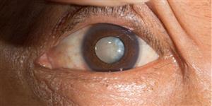
If you have a cataract, your lens has become cloudy, like the bottom lens in the illustration. It is like looking through a foggy or dusty car windshield. Things look blurry, hazy or less colorful with a cataract.

The definition of a cataract is a cloudy lens in the eye, whatever the cause may be. Here the cataract lens is compared to a natural clear lens.

The top lens is a clear, natural lens. The bottom lens shows clouding by cataract.

What Are the Symptoms of Cataracts?

Here are some vision changes you may notice if you have a cataract:

Dull or yellowed vision from cataracts.

Dull or yellow vision
from cataracts.

Blurry or dim vision from cataracts.

Blurry or dim vision is a symptom of cataracts.

Distortion or doubled images from cataracts.

Distortion or ghost images from cataracts.

See a simulation of what vision with cataract looks like.

What Causes Cataracts?

Aging is the most common cause. This is due to normal eye changes that happen starting around age 40. That is when normal proteins in the lens start to break down. This is what causes the lens to get cloudy. People over age 60 usually start to have some clouding of their lenses. However, vision problems may not happen until years later.

Other reasons you may get cataracts include:

Most age-related cataracts develop gradually. Other cataracts can develop more quickly, such as those in younger people or those in people with diabetes. Doctors cannot predict how quickly a person’s cataract will develop.

You may be able to slow down your development of cataracts.

Protecting your eyes from sunlight is the best way to do this. Wear sunglasses that screen out the sun’s ultraviolet (UV) light rays. You may also wear regular eyeglasses that have a clear, anti-UV coating. Talk with your eye doctor to learn more.